
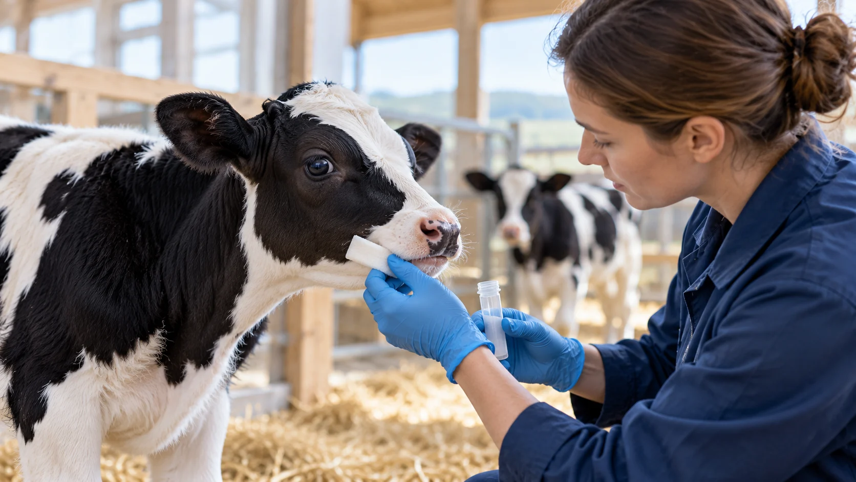
In short: Saliva is easy to collect repeatedly from cattle, so it is attractive for tracking infection. But three recent U.S. Department of Agriculture studies, two on H5N1 avian influenza and one on bovine tuberculosis, show that saliva is not always a reliable sample. In the calf study, nasal swabs detected infection in more animals than saliva did, and in the tuberculosis study neither saliva nor nasal swabs detected infection reliably. That is useful for researchers planning which samples to take and when.
This article describes published research. Oasis Diagnostics devices are for research use. The findings concern the samples in these studies, not the devices.
Key takeaways
- In experimentally infected calves fed H5N1-contaminated milk, nasal swabs were positive in all 4 of 4 calves in the first group, while saliva was positive in 2 of 4.
- In calves in a second group, all saliva samples were negative.
- In an experimental bovine tuberculosis study of 18 calves, M. bovis DNA was detected in saliva from a small number of animals, culturable bacteria were never recovered from saliva, and the authors concluded shedding was infrequent, intermittent and at very low levels.
- Saliva still earned a place in the sampling plan: it was collected from all animals with a non-invasive absorbent pad (Super•SAL or SuperSal) at regular intervals.
Study 1: H5N1 in dairy cattle
A 2025 Nature study experimentally infected Holstein heifers by an aerosol respiratory route and lactating cows by an intramammary route with H5N1 clade 2.3.4.4b. Samples included nasal, oropharyngeal, ocular and rectal swabs, blood, milk and saliva, taken daily for 7 days after inoculation and about every two days afterwards. Saliva was collected with an absorbent pad (Super•SAL).
In the aerosol-inoculated heifers, nasal, oropharyngeal, saliva and ocular samples sporadically had low cycle-threshold values (a measure of viral RNA). One heifer had viral RNA detected across several days in oropharyngeal swabs, ocular swabs or saliva. The paper’s headline findings are about the infection itself: the lactating cows developed mastitis, while the heifers showed no overt signs of illness beyond transient increases in nasal secretions.
Study 2: calves fed infected milk
A 2026 Nature Communications study fed calves unpasteurized milk from infected cows. Behaviour, respiratory effort and temperature were monitored and ocular, nasal and rectal swabs, blood and saliva were collected daily for 6 days. Saliva was collected with an absorbent pad (Super•SAL) before feeding each day.
| Group | Nasal swabs | Saliva |
|---|---|---|
| Group One (milk with virus, no antibody), 4 calves | PCR-positive in 4 of 4 calves, from 2–3 days after feeding | PCR-positive in 2 of 4 (one on day 1, one on day 3) |
| Group Two (milk with antibodies), 4 calves | Negative in 3 of 4 calves | Negative in 4 of 4 at all time points |
Serum and faecal swabs were negative in every calf in both groups. The study found that calves fed infected milk became infected, and that antibodies in the milk gave protection.
Study 3: bovine tuberculosis
A 2024 study in Veterinary Sciences looked for Mycobacterium bovis in body fluids of 18 calves during the first 8 weeks after experimental aerosol infection. Saliva was collected 7 days before infection and then weekly for 8 weeks using SuperSal devices, with nasal swabs and blood collected on the same schedule.
- M. bovis DNA was detected in saliva from a small number of calves at a few time points, and in nasal swabs from a small number of calves at weeks 5, 6 and 8.
- No culturable M. bovis was ever recovered from nasal swabs or saliva.
- No M. bovis DNA was found in blood.
- Every calf was shown to be infected by an interferon gamma release assay.
- Only one animal had both a PCR-positive nasal swab and saliva sample, at 8 weeks.
The authors write that bacterial shedding in nasal secretions or saliva was infrequent, intermittent and at very low levels, contrary to their hypothesis, and that identifying infected animals by detecting the bacterium would need alternative samples or assays.
What this means for sampling plans
- Use more than one sample type. In the calf study nasal swabs detected infection in more animals than saliva, and in the tuberculosis study neither saliva nor nasal swabs detected infection reliably.
- Sample repeatedly. Positives were sporadic, so single time points would have missed them. Saliva makes repeated sampling practical.
- Match the sample to the pathogen. Nasal swabs worked better for H5N1 in calves, and neither nasal swabs nor saliva worked well for early bovine TB.
- Plan for low-level detection. When shedding is intermittent, assay sensitivity matters.
- Report negative results. These studies show saliva’s limits clearly, which helps others design better studies.
Collection devices for animal studies
- Super•SAL: a universal collection kit validated for saliva and samples from multiple animal species.
- Super•SAL 2: collects up to about 1 mL with no added preservatives and a volume indicator.
- Micro•SAL: for small animals including cats, small dogs and piglets.
- Research that used Oasis devices
Sources
- Baker AL, et al. Dairy cows inoculated with highly pathogenic avian influenza virus H5N1. Nature. 2025. https://doi.org/10.1038/s41586-024-08166-6
- Sarlo Davila KM, et al. Susceptibility of calves fed unpasteurized milk from cows infected with highly pathogenic avian influenza H5N1. Nat Commun. 2026. https://doi.org/10.1038/s41467-026-76300-1
- Palmer MV, et al. Pathogen detection in early phases of experimental bovine tuberculosis. Vet Sci. 2024. https://doi.org/10.3390/vetsci11080357